
Analytics
This site is under construction.
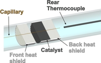
| Capillary-based in-situ sampling
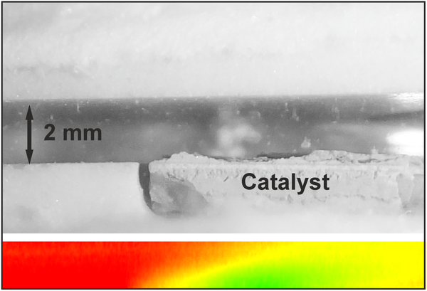
In-situ sampling of the gas-phase in a single channel of a monolithic honeycomb catalyst (tubular flow reactor capillary setup ) or above a catalytic plate (stagnation flow reactor) via a capillary technique for detection of spatially-resolved concentration profiles. Analysis of gas samples by FTIR, EI-MS and IMR-MS. In the channel of the catalytic monolith, also detection of temperature profiles possible. Measurement of gas-phase as well as surface temperature by thermocouple and optical fiber connected to a pyrometer, respectively. Ref.: |
|||
 |
 |
||
|
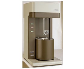
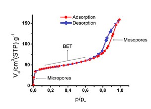
BET Analysis Gas adsorption is an important method for characterization of powder and porous materials. Our samples will be analysed on the BET surface analyser Belsorp-Mini. Nitrogen gas is applied to this technique. Measurements are carried out automatically after sample cells are set and the measurement data are analyzed with a dedicated software. Original dead volume measurements make it possible not to control refrigerant liquid level (LN2). Up to 3 samples can be measured independently and simultaneously. An adsorption isotherm can be obtained with nitrogen (N2), argon (Ar) and various other adsorptive gases. Ref.: |
|||
 |
 |
||
|
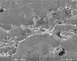
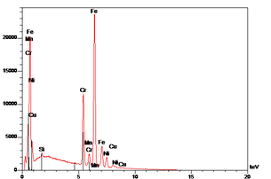
SEM Hitachi S570/EDX A scanning electron microscope (SEM) produces images of a sample by scanning it with a focused beam of electrons and gives information about the sample's surface topography. It can be achieved a magnification from 50 to 100 000. The resolution of SEM is about 1nm to 20 nm. Additional analysis with EDX (energy dispersive x-ray) is possible. |
|||
 |
 |
||
|
AAS Analysis Atomic Absorption Spectroscopy is a technique for determining the concentration of a particular metal element within a sample. The samples are analysed by the flame AAS Hitachi Z-6100. By utilising two optical pathways (sample / reference), the AAS guarantees a good baseline stability. Ref.: |
|||
 |
|||
|
PLIF (planar laser induced fluorescence) inside an optically accessible flow reactor In-situ spectroscopic method for 2d spatially resolved detection of excited species. Axial and radial concentration profiles of the excited species can be visualized simultaneously by PLIF above different catalytic surfaces from inside the reactor. After consideration of collisional quenching effects inside the probe volume absolute concentrations can be obtained. PLIF is a non-invasive method and possesses a high spatial and temporal resolution. Ref: |
|||
 |
|||